TL;DR: Proper preoperative testing is essential for safe and effective thyroid radiofrequency ablation (RFA). These tests confirm nodule suitability, reduce risks, and may be required for insurance coverage. Clinicians should follow a structured evaluation process before proceeding with ablation.
Key Pre-Op Tests for Thyroid RFA:
- High-Resolution Thyroid Ultrasound – Assesses nodule characteristics, size, and relationship to nearby structures.
- Fine-Needle Aspiration Biopsy – Confirms benign pathology before proceeding with ablation.
- Laboratory Tests – Evaluates thyroid function, blood count, and clotting factors to ensure procedural safety.
- Radionuclide Thyroid Scan – Determines whether the nodule is overactive (hot) or non-functioning (cold).
- Vocal Cord Function Assessment – Ensures vocal cords are functioning properly to avoid nerve-related complications.
- Additional Imaging (CT/MRI) – Used for large goiters or nodules extending into the chest for better anatomical visualization.
- Anesthesia and Nerve Block Planning – Ensures patient comfort and safety during the procedure.
By completing these essential tests, clinicians can confidently proceed with thyroid RFA, optimizing patient outcomes and procedural success.
For clinicians preparing a patient for thyroid radiofrequency ablation (RFA), a thorough preoperative RFA test regimen is essential. These pre-op evaluations ensure the thyroid nodule is a good candidate for ablation and can prevent potential procedural complications. This testing may also be necessary to ensure patients prequalify for insurance coverage, ensuring adequate reimbursement for qualifying cases.
The RFA testing process typically includes a range of imaging, lab work, and specialty exams. These tests verify that the nodule is benign, accessible, and that the patient can safely undergo the procedure.
In this blog, we’ll outline the key pre op tests for ablation of benign thyroid nodules. Continue reading to learn how clinicians effectively determine patient candidacy before the RFA procedure.
1. High-Resolution Thyroid Ultrasound Examination
Before undergoing RFA, every patient receives an ultrasound scan of the thyroid gland and neck to assess the nodule’s characteristics. All international guidelines emphasize a thorough ultrasound RFA medical test as a first step.
An initial ultrasound of the thyroid is necessary, not only for planning the RFA approach, but to identify features typically associated with malignancy. Such features include irregular margins, extrathyroidal extension, or abnormal lymph nodes.
Ultrasound can confirm:
- The nodule’s size (all three dimensions) and volume
- Nodule composition (solid, cystic, or mixed)
- Internal vascularity
- Relationship to nearby critical structures
Essentially, a nodule with benign ultrasound features provides confidence to move forward with ablation once other tests are cleared. The scan also serves as a baseline to measure how much the nodule shrinks after RFA.
2. Fine-Needle Aspiration Biopsy (Cytology Confirmation)
At present, RFA is only indicated for the treatment of benign thyroid nodules. As a result, it’s crucial to rule out potential malignancy before proceeding with ablation.
Fine-needle aspiration (FNA) cytology is the standard test before radiofrequency ablation to establish benign pathology. At present, clinical guidelines recommend two benign cytology results before proceeding with RFA. This minimizes the risk of inadvertently ablating a cancerous nodule that would be better treated surgically.
3. Laboratory Tests: Thyroid Function and Blood Work
Once a nodule is confirmed benign, patients undergo a panel of blood tests to check thyroid hormone levels and general health parameters.
The standard panel includes:
- Thyroid function tests (TFTs): Thyroid-stimulating hormone (TSH) plus free T3 and free T4 levels.
- Complete blood count (CBC): To ensure the patient has no underlying infection or hematological issues.
- Coagulation profile: Blood clotting tests (e.g., PT/INR, aPTT) to check bleeding risk.
According to international guidelines, thyroid function should be evaluated pre-procedure for every RFA patient. CBC and coagulation tests are also recommended to ensure there’s no anemia, thrombocytopenia, or coagulopathy that could complicate the procedure. If a patient is on anticoagulant or antiplatelet medications, those are usually paused in the days before ablation to reduce bleeding risk.
4. Radionuclide Thyroid Scan (for Hyperfunctioning Nodules)

A thyroid scintigraphy scan shows whether a nodule is “hot” (overactive) or “cold” (inactive) in terms of uptake. In practice, if a nodule is “hot” on scan, the patient may need beta-blockers or other measures before and during RFA to manage symptoms of hyperthyroidism.
After bloodwork, a patient’s levels might indicate that a nodule could be an autonomously functioning thyroid nodule (AFTN). Such nodules are still potentially candidates for RFA. However, gathering more information via nuclear medicine scans can help clinicians to guide management. For example, larger toxic nodules might require multiple ablation sessions to achieve volume reduction.
5. Vocal Cord Function Assessment (Laryngoscopy if Indicated)
The thyroid gland is located near the recurrent laryngeal nerves. Though RFA carries a low risk for nerve injury, standard practice is to document baseline vocal cord function pre-procedure.
Current guidelines recommend laryngoscopic examination before ablation in patients who have:
- Voice changes
- A history of neck or thyroid surgery
- Nodules very close to the trachea or nerve structures
For example, a patient with prior thyroid surgery might have an asymptomatic vocal cord palsy.
In routine cases without risk factors, a formal scope might not be required. At minimum, however, the clinician should perform a voice assessment for hoarseness before proceeding with RFA.
6. Additional Imaging if Needed (CT or MRI for Large Goiters)
In certain scenarios, specialized imaging of the neck and chest, such as a CT scan or MRI, can provide more detail on the thyroid nodule’s anatomy. This is necessary if a nodule is extremely large or extends into the chest. An intrathoracic thyroid extension can be better delineated with CT scan, for example.
CT or MRI can reveal the nodule’s relation to the windpipe, esophagus, and blood vessels. This is valuable information for a safe ablation approach for large goiters.
7. Anesthesia and Nerve Block Planning
Ensuring proper anesthesia is a critical step before radiofrequency ablation. All major guidelines recommend using local anesthetic (rather than general) for thyroid RFA. Local anesthetic is safer and allows real-time feedback from the patient.
Prior to starting ablation, the clinician performs a nerve block test of sorts. They inject lidocaine around the thyroid capsule to numb the area and block pain signals from the nerves in that region. This step is sometimes informally referred to as a “nerve block test before radiofrequency ablation.” It ensures that the patient is comfortable and that critical nerves are insulated by anesthetic fluid.
Effective RFA medical test dosing of local anesthetic is confirmed when the patient reports neck numbness and tolerates the probe position without pain. In some cases, light conscious sedation or an anxiolytic is given as well. Most clinicians prefer to avoid deep sedation so the patient can alert them to any symptoms during the procedure itself.
Once the area is adequately numbed and all other tests above are clear, clinicians may proceed with the ablation.
Incorporate RFA Into Your Clinical Practice
Each RFA test plays a role in reducing risk and optimizing outcomes. By completing these pre op tests for ablation, clinicians can proceed confidently, knowing they have assessed the nodule from all angles. Proper patient selection and preparation sets the stage for a smooth RFA session and the best possible result for the patient.
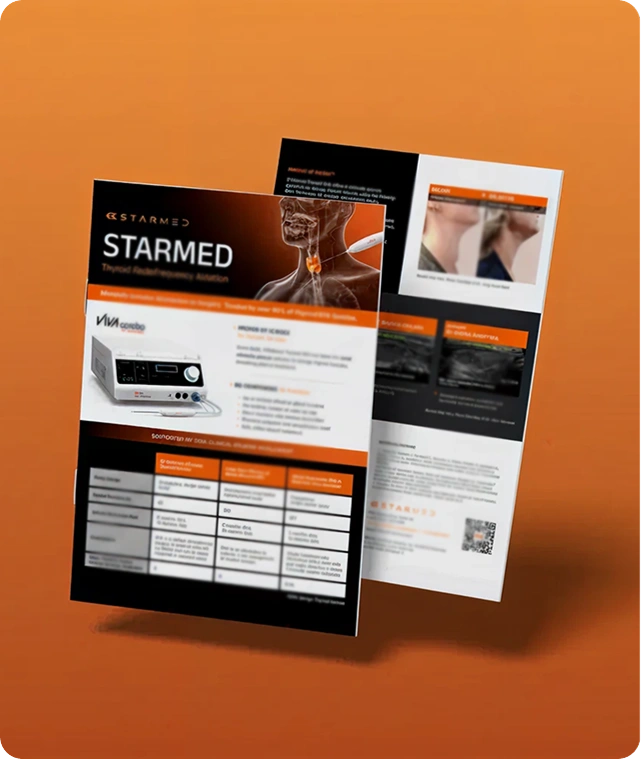
Mastering the thyroid RFA procedure begins with comprehensive training and support for clinicians. Learn more about Thyroid RFA and discover how STARMED America can help you implement the procedure into your practice.